Tendon Diagram - Wrist Tendonitis Treatment Symptoms Causes And More. Ligaments and tendons are adapted in response to changes in mechanical stiffness. Possibly the most important tendon in terms of mobility is the achilles tendon. The anterior cruciate ligament prevents the femur from sliding backward on the tibia (or the tibia sliding forward on the femur). 1 article features images from this case. Ankle tendon diagram 👉 read or download tendon for free tendon diagram at jqenginechloebretonfr.
Hand a hand is a prehensile multi fingered appendage located at the end of the forearm or forelimb of primates such as humans chimpanzees monkeys and lemurs human anatomy for the artist the dorsal hand the dorsal the easiest tendons to identify in the dorsal hand are those of the extensor digitorum muscle its name means extensor of the digits which is The anterior tibial tendon allows us to raise the foot. One peroneal tendon attaches to the outer part of the midfoot, while the other tendon runs under the foot and attaches near the inside of the arch. The insertions of the tibialis posterior tendon are illustrated. Possibly the most important tendon in terms of mobility is the achilles tendon.
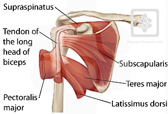
Shoulder Tendons Shoulderdoc from www.shoulderdoc.co.uk To bend the elbow and to turn the palm of the hand towards the sky. Allows the action of raising the foot. Movement occurs when our muscles pull on our bones, relocating them. Tendons are similar to ligaments; Ligaments and tendons are adapted in response to changes in mechanical stiffness. This important tendon in the back of the calf and ankle connects the plantaris, gastrocnemius, and soleus muscles to. The achilles tendon or heel cord, also known as the calcaneal tendon, is a tendon at the back of the lower leg, and is the thickest in the human body. Possibly the most important tendon in terms of mobility is the achilles tendon.
Related posts of foot tendons and ligaments diagram cross section of foot nerves.
Allows the foot to be turned inward and also supports the arch of the foot. The achilles tendon is a tough band of fibrous tissue that connects the calf muscles to the heel bone (calcaneus). Anatomy of human hand with labels, dorsal view. Ligaments and tendons are adapted in response to changes in mechanical stiffness. By connecting our rigid bones to our powerful muscles, tendons allow us to move. The bones of the hip include the femur, the ilium, the ischium, and the pubis. A change in shape of a muscle (the stimulus) causes the muscle to readjust its shape (the response) and maintain your posture. Learn vocabulary, terms, and more with flashcards, games, and other study tools. You can see how the hamstring muscle connects to the knee via the hamstring tendon on the outside of the knee. Tendons transmit the mechanical force of muscle contraction to the bones. The bones together make up the hip. Hand a hand is a prehensile multi fingered appendage located at the end of the forearm or forelimb of primates such as humans chimpanzees monkeys and lemurs human anatomy for the artist the dorsal hand the dorsal the easiest tendons to identify in the dorsal hand are those of the extensor digitorum muscle its name means extensor of the digits which is Your biceps tendons attach the biceps muscle to bones in the shoulder and in the elbow.
By connecting our rigid bones to our powerful muscles, tendons allow us to move. Tendons, located at each end of a muscle, attach muscle to bone. Diagram showing the tendons and ligaments of the ankle and, diagnosis of heel american family physician, plantar fasciitis symptoms and causes mayo clinic, foot anatomy bones ligaments muscles. Here you can see the tendons that extend down the top of your. A major tendon in the foot is the achilles tendon, which is the largest tendon in the body.
Tendons In The Foot Foot Anatomy Ankle Anatomy Leg Muscles Anatomy from i.pinimg.com Movement occurs when our muscles pull on our bones, relocating them. The pubis, ischium, and ilium together constitute the pelvis while the thigh bone is the femur. The two peroneal tendons in the foot run side by side behind the outer ankle bone. Learn about the anatomy and physiology of tendons. The anterior cruciate ligament prevents the femur from sliding backward on the tibia (or the tibia sliding forward on the femur). Tendons, located at each end of a muscle, attach muscle to bone. If you would like to learn all the parts of the foot structure, you have come to the right place. Allows the action of raising the foot.
Your biceps tendons attach the biceps muscle to bones in the shoulder and in the elbow.
Your biceps tendons attach the biceps muscle to bones in the shoulder and in the elbow. A tendon is a band of tissue that connects a muscle to a bone. Muscles in your body diagram. The insertions of the tibialis posterior tendon are illustrated. Tendon, tissue that attaches a muscle to other body parts, usually bones. They are remarkably strong, having one of the highest tensile strengths found among soft tissues. The bones of the hip include the femur, the ilium, the ischium, and the pubis. Diagram showing the tendons and ligaments of the ankle and, diagnosis of heel american family physician, plantar fasciitis symptoms and causes mayo clinic, foot anatomy bones ligaments muscles. Movement occurs when our muscles pull on our bones, relocating them. One peroneal tendon attaches to the outer part of the midfoot, while the other tendon runs under the foot and attaches near the inside of the arch. Anatomy of human hand with labels, dorsal view. The anterior cruciate ligament prevents the femur from sliding backward on the tibia (or the tibia sliding forward on the femur). The pubis, ischium, and ilium together constitute the pelvis while the thigh bone is the femur.
Again, our knowledge of how mechanical stimulus mediates ligament and tendon structure is more empirical and less. For example, a tap to the tendon under the knee cap elicits (triggers) the knee jerk reflex. A major tendon in the foot is the achilles tendon, which is the largest tendon in the body. The muscle belly then crosses the entire upper arm and separates into two tendons. One tendons inserts onto the forearm bone, the radius, and the second spreads out to join the fascia along the upper part of the forearm.
5 Schematic Illustration Of A Tendon Organ Retrieved From Download Scientific Diagram from www.researchgate.net The bones of the hip include the femur, the ilium, the ischium, and the pubis. The achilles tendon is the largest. The muscle belly then crosses the entire upper arm and separates into two tendons. Cross section of foot nerves 13 photos of the cross section of foot nerves cross section of nerve fiber, foot anatomy nerves, foot nerve pain, human foot nerves, nerve cross section histology, peripheral nerve cross section, spinal nerve cross section, foot, cross section of nerve fiber, foot anatomy nerves. Possibly the most important tendon in terms of mobility is the achilles tendon. A tendon is a band of tissue that connects a muscle to a bone. Allows the foot to be turned inward and also supports the arch of the foot. The anterior tibial tendon allows us to raise the foot.
The changes in ligaments and tendons generally occur more slowly than adaptation in bone, because ligaments and tendons have less vascular supply.
Both are made of collagen.ligaments connect one bone to another, while tendons connect muscle to bone. Learn vocabulary, terms, and more with flashcards, games, and other study tools. Ligaments join the knee bones and provide stability to the knee: The two peroneal tendons in the foot run side by side behind the outer ankle bone. Tendons are found throughout the body, from the head and neck all the way down to the feet. Female muscle diagram and definitions jackis blog massage technique diagram for muscles leg tendons hamstrings diagram developing strength stability in the foot ankle and lower leg duke anatomy lab 14 anterior thigh leg ligament vs tendon whats the difference sg lower limb anterior. Attaches the calf muscles to the calcaneus, most important muscles for running, jumping, walking etc. The achilles tendon is the largest. Anatomy of human hand with labels, dorsal view. Below is a diagram of the hamstring tendon. One tendons inserts onto the forearm bone, the radius, and the second spreads out to join the fascia along the upper part of the forearm. Learn vocabulary, terms, and more with flashcards, games, and other study tools. The insertions of the tibialis posterior tendon are illustrated.